
Coccidia is a species of protozoa organisms.
These are parasites of epithelial cells, which are the cells that recover surfaces, like skin, or in this specific case, GI tissues.
Of the 12 species of Coccidia associated with rabbits, it is not abnormal to find that one single rabbit has two or more species at the same time. Different coccidia species could affect different systems.
Rabbits have two common ways of getting infested:
1. By eating: wild fresh plants like grass can contain the Oocytes (eggs) of coccidian species.
2. By other rabbits: As a GI infestation, coccidia oocytes are expelled through feaces.
Oocytes can live up to a year outside a body and once certain requirements are met these hatch in the span of two days.
These faeces can be ingested or, by just the touch with food and water, release the oocytes, which are later on ingested by other rabbits.
Coccidia and the oocytes normally present themselves in the rabbit droppings, thus it can be diagnosed through faecal analysis, but as most often times coccidial infestations are asymptomatic, rabbits are not tested.
Asymptomatism does not mean rabbits can’t further develop the disease, which usually occurs due to lower defences because of stress or disease.
Coccidian infestations inhibit or hypertrophy the cells they infest, leading to nutrient malabsorption, anaemia, hypoprotenemia, and dehydration, amongst other.
An intestinal coccidiosis can develop general sickness symptoms (non-specific symptoms) like lack of appetite and depression, and the infestation itself causes weight loss, abdominal pain and diarrhoea, possibly haemorrhagic, due to the damage caused to the intestinal walls.
Sadly, intestinal infestations are usually diagnosed post mortem once the rabbit has gone through a weight loss cycle. This cycle ends once the rabbit reaches 20% of its needed bodymass, causing sock with convulsions and/or paralysis.
Intestinal coccidiosis mainly affect rabbits from 6 weeks to 5 months old, and is attributed to stress and/or a weak immune response.
Young rabbits can be very susceptible to Coccidian species, Eimeria stiedai being one of them. This genus causes damage by obstructing the hepatic bile ducts and causing liver fibrosis (the liver tissue gets thick or scarred) leading to hepatic damage. It develops into diarrhoea, weight loss, jaundice (due to the liver damage), ascites (liquid retention in the abdomen), and slowed growth in younger individuals.
Unlike other mammals, treatment is life-long and it is not definitive as the disease commonly relapses after a week or two, and only infestations caught early have a chance of full recovery.
Treatment does not guarantee that the rabbit will not succumb to the disease, and Coccidia can slowly become resistant of the preventive medicine that can be given to rabbits, usually specific anti-parasite for coccidia.
If you want to learn more about Coccidia and coccidiosis you can see:
The Unusual Pet Vets’ PDF about Coccidia.
MediRabbit’s extensive information sheet on Protozoal enteritis caused by Coccidiosis.

A B
Cystoisospora species.
C
Coccidia oocyst with multiple sporocysts.
D
large brown coccidia.
E
Eimeria oocyst.
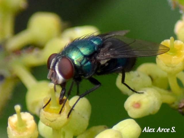

Myiasis is the infestation of fly larvae in tissue.
It is common in animals that are either very dirty or live in very dirty conditions.
For rabbits, a myiasis is considered an emergency and the longer the rabbit has the larvae within its body, the higher the chances of death.
Every fly is capable of causing flystrike, but the Calliphoridae family is notable as they don’t require an open wound to begin infesting. They are also known for using other flies to carry their eggs.
Myiasis occurs when flies, attracted by smell (from urine or faeces) or open wounds lay their eggs to grow on the tissue.
These hatch into larvae that start feeding on the flesh of the infested animal, opening a bigger wound and thus attracting more flies.
Despite this, myisis infestations tend to be quite clean, because the larvae tend to only go for the rotting flesh rather than healthy flesh.
Lucilia cuprina’s maggots, another fly from the Calliphoridae family, expel ammonia as a waste product from eating tissue which intoxicates the host.
These are mainly found on Australia.
Preventive action includes ensuring the rabbit and their areas are clean.
A rabbit may be dirty due to an incapability of cleaning themselves (Obesity, Arthritis), or to sickness (Diarrhoea, Urinary issues).
It is also important to keep any open wound clean, dry, and sterile.
Treatment usually involves active removal of the maggots from the wound with a medicinal larval pesticide. It is important to consider not all pesticides work for all animals, and rabbits could develop hypersensitivity reactions or toxic shock to those aimed at dogs and cats.
These pesticides tend to be quite harsh on the wound and animals often times struggle in mild pain and discomfort.
If the debridement is done in especially soft tissue like the anus, and as a general well-being practice, you should provide your patient with mild painkillers.
To learn more about flystrike in rabbits, you can visit:
Rabbit Welfare Association and Fund’s information about flystrike.
Bunny Lady’s article about flystrike and how to prevent it.
To learn more about flystrike in general, or the Calliphoridae family, you can see:
Wikipedia’s article about myiasis (contains harsh images).
Wikipedia’s article about the Calliphoridae family.
Wikipedia’s article about Lucilia cuprina or the Australian sheep bowfly.
E. Cuniculi is known as a spore-forming parasitic fungus, where Encephalito- from encephalitis, means inflammation of the brain, and -zoon from zooid, means organism, and where cuniculi is the plural of cuniculus, meaning small conduits, indicating the way this pathogen reproduces.
E.C. can be also found in the rabbit’s kidneys due to the way it gets expelled from the body, so kidney damage and failure has been observed.
E. Cuniculi is known to develop asymptomatic infestations, triggering based on immune depression by stress or primary disease.
It can also be transmitted by another rabbit through urine, thus ingestion of contaminated water or food is the most likely cause.
E.C. is treatable, but damage in the ear or the brain could leave the rabbit with headtilt for the rest of their lives.
If caught early, the rabbit can recover from the symptoms, including headtilt.
Encephalitozoon Cuniculi and headtilt come hand in hand, although E. Cuniculi is not the only cause for headtilt.
Headtilt occurs when the inner ear of the rabbit gets infected and/or damaged (Peripheral) or when a pathogen enters the part of the brain that connects with the ear (Central).
In both cases, the rabbit loses balance and their sense of direction, their eyes can become damaged, causing them to turn their head to correct the position the brain thinks it is in.
Damage on the inner ear may occur due to many things, such as bacterial infections that expanded from the respiratory tract, or due to trauma.
Neurological damage can be caused by bacterial infections or by E. Cuniculi, but other symptoms are developed before headtilt, such as erratic movement, seizures, paralysis, or lack of appetite amongst others.
Bacterial headtilt can be reversible depending of the stage of infection, but headtilt due to trauma or parasitic infestation are not completely reversible, although it has been observed that the heads do straighten a bit after parasitic infestations are cured.
Strokes due to E. Cuniculi are easily recognisable, as the rabbit completely loses control and starts spinning over itself.
You can learn more about E. Cuniculi and headtilt from our sources below:
Wikipedia’s entry on E. Cuniculi.
Rabbit Welfare Association and Fund’s information sheet about E. Cuniculi.
The Educated Rabbit’s extensive information sheet about headtilt and its causes.


A
Swollen upper eyelid with edema.
B
Conjuntivitis with lacrimation.
C
Pseudotumour with crust emerging from a Myxoma on the upper lips.
Myxomatosis is a viral disease only affecting Lagomorphs.
Its origins trace the virus to South and North America.
For native american lagomorphs, the disease usually causes nodules as the sole symptom, in European rabbits though, the disease is fatal.
The Californian and Mexican strain is the most virulent, whilst the South and Central American one is slightly less fatal.
The current European and Australian strain is the least fatal.
The main way of transmission is through insect bites, especially fleas and mosquitoes that have already fed from a sick rabbit.
A secondary transmission route are body secretions, like snot from the nose or secretions from the reproductory organs.
This makes all rabbits, regardless of where they live, prone to get infected.
House rabbits are less prone to infection as they are not in possible constant contact with wild rabbits, although it still can enter the house through shoes and clothes.
Rabbits that have garden time or live outside need to be protected further by ensuring no wild rabbit can enter their land or play area.
You can also protect your rabbit by vaccinating them.
Vaccination does not guarantee that the rabbit will not get infected, but it greatly improves the chance of survival.
To ensure recovery a vet will provide palliative care.
This may depend on how advanced and symptomatic the disease is.
The usual causes of death are based on the symptoms, some of which can be read on The Blue Cross’ page sourced below, and include:
Extensive swelling of the entire body, including the eyes, which can lead to blindness. This swelling may also close the respiratory tract, making it harder or impossible for the rabbit to breath.
Myxoma tumours, wounds and lesions.
Discharge from eyes, nose, and other orifices. This discharge is mucopurulent in most strains, but some can develop a clear-looking one. The discharge can also be ingested and breathed, with a chance of developing into pneumonia.
Palliative care may look like:
1. IV Fluids – especially if the rabbit can’t ingest due to the myxoma tumours. Based on dehydration levels you may have to treat the rabbit as hypovolaemic with high infusion rates (120mL/kg/d).
2. Syringe feeding or Nasopharyngeal (or equivalent) tube feeding, again, if the rabbit can’t physically ingest.
3. Disinfection of myxoma wounds and lesions.
4. O2 supply or assisted breathing though intubation or otherwise.
Due to the systemic inflammation throughout the body, we can provide some NSAIDs like:
– Medetomidine (0.25mg/kg PO/IM). Shouldn’t be used if heart, kidney or liver issues suspected. To prevent kidney or liver damage, ensure proper hydration previous to administration.
-Carprofen (1-2mg/kg IV/SC). Can be used alongside buprenorphine in multimodal analgesia.
-Meloxicam/Metacam/Meloxidyl (0.1-0.2mg/kg PO). Rabbits have high tolerance, and if strictly necessary, you could increase the dose up to 1.5mg/kg.
If you want to learn more about myxomatosis you can read:
The Rabbit Welfare Association and Fund’s brief on Rabbits and Myxomatosis.
The Blue Cross’ information sheet about What is Myxomatosis.
Wikipedia’s article in Myxomatosis.
You can also check this “Management of Rabbits” page from MSD Manual for more information on general veterinary care knowledge.
Rabbits, alike many other animals, are prone to be infested by parasites, whether these are found in or outside of their body.
We have already talked about some of them, like Coccidia, E. Cuniculi, and Myisis, three horrible infestations with a variety of symptoms.
Now we must learn about other less lethal, but still harmful, parasites.
Skin parasites (ectoparasites) include external parasites found in fur or skin:
Mites like Cheyletiella affect dogs, cats, and rabbits, and big infestations are usually caused because the rabbit is immune deficient, meaning it’s secondary to an already ongoing issue.
There are also mites that are specialised to live inside the ears, but these are harder to diagnose because bacterial and fungal infections may develop similar symptoms, thus it is recommended for a vet to check under a microscope.
Ticks and fleas also affect rabbits.
Fleas can be dealt with by thoroughly cleaning the environment and applying the necessary pest control, of course this must be done after removing the rabbit, who also has to be cleansed. The most important part of flea removal is ensuring their eggs and larvae are gone, as those are the usual suspects when consequent infections occur.
There are fleas specialised in rabbits and they usually target the ears.
Flea allergy may also be developed, just like any other animal.
Ticks can also be a big part of rabbit health, and big tick infestations can easily and quickly debilitate rabbits by anaemia.
It is important to check yourself and especially other animals that can co-inhabit with the rabbit if these go outside often.
Cats and dogs can bring ectoparasites into the house easily.
Remember to check your rabbit often if they can access the garden or live outside.
Myiasis occurs when a fly deposits its eggs in the rabbit flesh, the larvae hatch and start feeding on the rabbit. This is also considered a parasitic infestation.
Mosquitoes are a matter of worry due to how harder they are to control compared to the others on the list. They are well known for carrying blood borne RHVD.
There are other parasites that affect rabbits from within:
E. Cuniculi is one of them, infecting the brain and other organs and causing a plethora of issues like paralysis, seizures, and headtilt.
Baylisascaris, a roundworm found in raccoons, can infect the eyes and the brain alongside the rest of the organs and has similar symptoms to E. Cuniculi.
Coccidia are parasites infecting the GI tract, they affect the lining of the intestines, causing weight loss and diarrhoea among other things. There is also a second type of Coccidia that infects the liver causing hepatic damage.
Rabbits are also prone to pinworms, tapeworms, and other round worms.
Vets are able to give you anti-parasite drops or sprays that allow for protection and diagnosis. If you buy these yourself from a pet shop please remember: Most of these sprays made for dogs and cats are toxic for rabbits.
In case of parasitic infection, other preparation and care must be made with the veterinary expert.
For your general internal parasites, panacure is quite effective, and a tri-monthly cure can be done if necessary.
For some bacterial intestinal parasites, antibiotics can be shyly used, always considering that some antibiotics are toxic for rabbits and providing rabbit-specific probiotics and prebiotics
Rabbits can be treated with some medicines for external parasites too, including Ivermectine and Selamectine, which also work for some internal parasites too.
I personally use Selamectine (Stronghold).
Be wary that some anti-parasite meds are toxic to rabbits too.
If you want to learn more about parasites you can read:
The Rabbit Welfare Association and Fund’s page on Skin and Fur Parasites.
Exotic Pet Vet’s information sheet about Parasites of Rabbits.


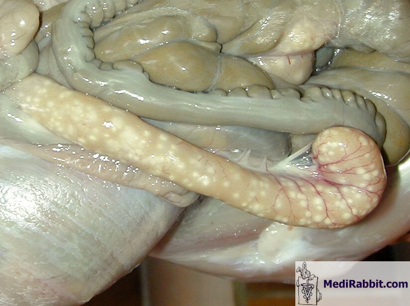
A
Post-mortem side view.
B
Bleeding from the nose.
C
Liver necrosis.
D
Close-up of a liver lobe.
(More detailed information in the source)
Rabbit Viral Haemorrhagic Disease or RHVD are two strains of viral hepatitis only affecting rabbits, with a mortality of 70-100%.
Some strains also affect hares and cottontails.
Infection symptoms include liver necrosis and coagulation in most blood vessels, worsening the necrosis and reducing the blood and oxygen supply to other organs. Blocked vessels may rupture due to the pressure, creating internal bleeding and external bleeding through orifices like the nose.
Most RHVD strains only have internal symptoms, thus showing no clear signs of disease apart from not-specific symptoms, to the point where some rabbits die seemingly by random chance, and rarely catching the disease on post-mortem revisions.
This is due to a peracute case, where rabbits die extremely fast and prematurely.
In acute cases bunnies show general sickness symptoms alongside high fevers, bloody discharge from orifices and in poop and urine. Lethargy, coma and convulsions are common before death, and rabbits with these symptoms die within 12 to 36 hours after developing a fever.
Chronicity is a common symptom of RHVD 2, other symptoms including general sickness symptoms, anorexia, and jaundice (yellowing of the eyes and other tissues) due to high levels of bilirubin due to liver failure. GI dilation, arrhythmia and heart murmurs, and neurological symptoms may develop. Death comes within 1 to 2 weeks and usually occurs due to liver failure.
Rabbits can also be asymptomatic, and due to the incubation period, especially of RHVD2 (3-5 days), the rabbit can infect other rabbits without prior notice.
The good news is that there are very effective vaccines, thus it is very recommended for you to vaccinate your rabbits accordingly for both strains of RHVD.
Some vaccines include Myxo + RHVD 1+2.
Recently, a new strain of RHVD was discovered in certain European countries. Due to information being kept from most people, I am yet to know more about it, but apparently there is a vaccine.
If you want to learn more about RHVD, you can read:
The Rabbit Welfare Association and Fund’s information sheet about RHVD.
Wikipedia’s article on Rabbit Haemorrhagic Disease.
Snuffles is the generation of mucous in nose and eyes due to a bacterial infection within the sinuses and tear ducts.
This mucus may also be accompanied by sneezing and wheezing, which means the infection has reached the later stages.
It is not uncommon to see general sickness symptoms alongside drooling (due to mouth-breathing), and dirty, matted paws, as they try to clean the mucous from their face.
Skin sores may also develop due to the constant wiping and liquid within the nose and eyes areas.
Rabbits with dental diseases are prone to developing snuffles due to how close the tooth roots are to the tear ducts. Malocclusion can also create issues as the deformity causes the ducts to close, creating a special environment for bacteria to grow.
If the infection reaches the ears, it may cause reversible headtilt. Reversible meaning that partial or complete tilt may be cured. Regardless, in very advanced infections where headtilt is severe, it may become hard to reverse the tilt.
In mild cases, the symptoms can be easily controlled and the rabbit can heal. In grave cases, rabbits can develop pneumonia, which greatly increases mortality.
Sadly, these infections are hard to eradicate, and can only be prevented with proper care and controlled with medication, thus it is not uncommon to find that a rabbit that has previously been infected has developed snuffles again.
Ensuring your rabbit isn’t stressed and has a healthy immune system is a great way of reducing the chance infection and reducing relapses.
The bacteria commonly causing infection are Pasteurella spp. and Staphylococcus spp., but other bacteria like Bordatella Bronchiseptica (also affecting humans and dogs) and Pseudomonas can also infect your rabbits.
If you want to learn more about snuffles, you can always visit:
The Rabbit Lady’s page on Snuffles in Rabbits.
Bishops Stortford Veterinary Hospital’s facts list on Snuffles.
The Rabbit Welfare Association and Fund’s information sheet about Rabbit Snuffles.


Also called pseudotuberculosis, yersiniosis is a rare disease that usually affects adult animals.
The visible symptoms show as the rabbit eats less to the point of stopping, which drops their weight rapidly. Death follows soon after.
Droppings become hard and sticky, and their size decreases as food intake decreases.
When palpating, the belly is soft and you may be able to feel the content of the abdominal cavity decrease.
Birds and rodents normally carry the disease, these may include guinea pigs, so rabbits cohabitating with these animals can become infected. This is why cleanliness is key for prevention.
Wild rabbits can also become infected.
Hares are very sensitive and yersiniosis tends to be a common cause of death in some countries.
The bacteria can be found in both sick and healthy individuals as well as the general environment if in contact with faeces of an infected individual.
With a survival span of a year, the bacteria can reproduce at ranges of 4º C to 10ºC, which may explain the infection rates of rabbits in winter.
With high zoonosis, especially to children and immunosuppressed patients, it causes symptoms similar to appendicitis.
Both animals and humans can be affected by generalised infections, severe septicemia or localised infections in lungs or eyes.
The rabbit may show symptoms after the approximate 15 day incubation period.
Septicemic infections kill within 24 to 48 hours, whilst chronic forms could kill after 2 to 3 months, although these latter are commonly survivable.
The bacteria reaches the intestine and multiplies, thereafter it infects the lymphatic nodules.
Once the previous stage is reached, the liver is affected, which begins the septicemia.
With the other forms, the rabbit may show general sickness symptoms including exhaustion. Diarrhoea may also develop.
Yersiniosis can also target kidneys and lungs.
During palpation, you may be able to feel the hypertrophied
(large in size, sometimes swollen and hard) lymphatic nodules. The liver will have hard nodules too.
Whilst palpating you may find that the spleen may have grown in size (up to three times).
If there is exploratory surgery, white or yellow nodules (spots) may be seen in the ceacum, kidneys, and spleen, the intestine can present necrotic regions.
A diagnosis can be obtained by doing a biopsy of the affected organs.
If you want to learn more about yersiniosis you can visit (Careful, strong images):
Medirabbit’s page on rabbit tuberculosis.
Frances Harcourt-Brown’s image of the white/yellow nodules in the organs.

Create an account to use certain features and to register as a shelter.